
Shine On Hemangiosarcoma Project
|
||
| ||
Hemangiosarcoma Basics • Hemangiosarcoma in dogs originates from cells in the bone marrow that help in the formation of • Hemangiosarcoma cells travel throughout the body, increasing the risk that the disease will • There appears to be some risk for hemangiosarcoma associated with “being a dog,” and the risk • There are no known methods for effective prevention of hemangiosarcoma. Altering lifestyle • Hemangiosarcoma is almost always a fatal disease although its progression can be unpredictable. • The major goal of treatment is to prevent or delay a terminal bleeding episode. The most • Many other treatments are available for hemangiosarcoma, but none is shown to provide | ||
Studying Cancer in Dogs as a Path Towards a World Where We No Longer Fear
|


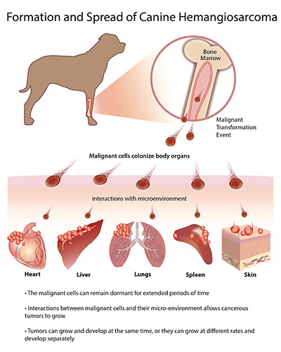 | Figure 1. Hemangiosarcoma appears to arise from bone marrow cells that aid in the formation of blood vessels. A transformation event can occur inside the bone marrow, or outside the bone marrow as these cells travel throughout the body to reach sites where new blood vessels are being made. As they circulate, the transformed cells can set up residence in multiple organs like the heart, liver, lungs, spleen, and skin. The cells can lie dormant for long periods of time in each of these sites, until a critical event takes place that leads to the formation of a tumor. One or more tumors can grow simultaneously, creating the patterns of metastasis seen in dogs with hemangiosarcoma. However, it also is possible for tumors to spread by cells moving from one site to another after the initial tumor forms. |
Hemangiosarcoma is a cancer that does not cause pain or discomfort. It can develop slowly without interfering with normal body functions and without obvious clinical signs. Tumor cells retain the ability to reside in and possibly form blood vessels, but, unlike normal blood vessels, the tumor cells inhabit and createmalformed vessels where blood tends to pool and clot. Eventually, these clots obstruct the vessels, preventing fresh blood and nutrients from reaching the tumor cells and causing some of them to die. Due to this cell death, the tumor develops ruptures where blood escapes into the abdomen, heart sac, chest, or subcutaneous space, depending on the location of the tumor. While substantial blood loss can lead to signs of anemia, such as pale gums, weakness, and lethargy, the signs may be subtle and can resolve as dogs reabsorb the blood components and make new blood cells, which is one reason why hemangiosarcoma almost always goes undiagnosed until the late stages of disease. By the time the cancer is diagnosed, it is almost certainly present in other sites, even though metastases may not be visible. Nonetheless, the eventual outcome for most patients is rupture of a tumor, with acute, severe blood loss, collapse, shock, and death.
How is hemangiosarcoma diagnosed in dogs?
The first step to diagnose hemangiosarcoma in dogs showing clinical signs is a complete and thorough physical exam, which may identify a mass. If there is any reason to suspect hemangiosarcoma, the next step is to conduct imaging tests, such as ultrasound or radiographs (x-rays). If the presence of a mass is confirmed, a biopsy, where a sample of the affected tissue or the entire mass is removed and the material is examined by a pathologist, is needed to definitively diagnose hemangiosarcoma. At this time, there are no readily available, effective tests to diagnose hemangiosarcoma before clinical signs appear. It is unclear if adding imaging tests to routine well health exams is helpful to diagnose developing tumors. Careful analysis of blood samples by an experienced pathologist may hint at the presence of bleeding episodes and blood vessel abnormalities that are suggestive of hemangiosarcoma. However, this method is neither sensitive nor specific to confirm the diagnosis.
If a diagnosis of hemangiosarcoma is suspected or confirmed, the next step is to determine whether the tumor has spread to other areas (called staging). This routinely includes basic blood and urine tests, chest x-rays, and ultrasound examination of the abdomen and potentially the heart. These tests are relatively sensitive. For example, ultrasound of the heart is able to identify the presence of a tumor in the heart 65-90% of the time. However, artifacts can obscure the interpretation of any of these tests. Advanced imaging modalities such as computed tomography (CT) and positron emission tomography scans (PET-CT) are being used more commonly, and appear to be more sensitive than conventional x-rays and ultrasound.
How is hemangiosarcoma treated?
As stated earlier, hemangiosarcoma in dogs is almost always fatal. Therefore, the principal goal of treatment is not necessarily to achieve a cure, but rather to slow down or delay the spread of the disease and to prevent or delay the occurrence of life-threatening bleeding episodes. This is why surgery to remove any visible tumor mass may be recommended for hemangiosarcoma patients whose condition is otherwise stable even if there is widespread metastasis.
Without treatment, most dogs diagnosed with hemangiosarcoma will die within one to two weeks, although some can survive for several months. For dogs with hemangiosarcoma of the spleen treated with surgery, the median survival time (that is, the length of time when half of dogs receiving this treatment would remain alive) is one to three months. Adding chemotherapy to the treatment, using protocols that include the drug doxorubicin given repeatedly at two to three-week intervals, or possibly a combination of multiple drugs given daily, increases the median survival time to four to six months, making the combination of surgery and chemotherapy the preferred and most effective treatment available for this disease. Hemangiosarcoma cells in most dogs inevitably develop resistance to chemotherapy, so only 10-15% of dogs diagnosed with hemangiosarcoma of the spleen will be alive one year or longer after their diagnosis. Studies performed in the last two decades using new combinations of old drugs, immunotherapy, and new drugs, have shown no benefit to improve survival for dogs with hemangiosarcoma of the spleen as compared to conventional surgery and chemotherapy. Survival time estimates for tumors located in other organs have more uncertainty, but generally the prognosis for tumors that involve the heart, liver, and other internal organs is worse than for tumors of the spleen, and the prognosis for tumors that are localized to the skin is better than for tumors of the spleen.
Alternative and complementary approaches continue to gain popularity in the search for a cure for hemangiosarcoma, but any publicity attributing curative power to a drug after an anecdotal response should be viewed with extreme caution. As noted above, some dogs with hemangiosarcoma will live a year or longer without any evidence of disease. In rare instances dogs will live for several years without disease recurrence. This is almost certainly due to the behavior of the tumors themselves; that is, hemangiosarcomas vary greatly, and some will show extremely slow disease progression regardless of the therapy used. Therefore, we strongly recommend options and treatment based on objective data and not on anecdotal information that creates false hope and unrealistic expectations in both pet owners and their veterinarians.
Hemangiosarcoma is a devastating disease. And while there is no cure, our research is showing promising results in the areas of treatments and early detection. This promise gives us hope that we are indeed closer to creating a world where we no longer have to fear cancer.
Note: This is an edited version of an article published in The Alpenhorn (The Official Publication of the Bernese Mountain Dog Club of America), Fall issue of 2017, pp. 64-68.